

Anatomical details
The anatomy of the 100% operable female reproductive system:
-
Uterus
-
Bladder
-
Round Ligament
-
Fallopian Tube
-
Ovary
-
Uterine Artery
-
Ureter
-
Suspensory ligament


Institutional Robotic Courses
In collaboration with Cyber Robotics, a Robot-Assisted Hysterectomy workshop was conducted in Mexico City, offering surgeons the opportunity to enhance their skills in a controlled training environment.
Participants followed a step-by-step surgical workflow using a high-fidelity simulation model that accurately replicates the female reproductive system, enabling standardized and reproducible robotic training.

Anatomical details
Includes a uterus similar to the hysterectomy model with 8 myomas:
-
Basic level (anterior myomas): M1 of 30mm (FIGO 5) and M2 of 21mm (FIGO 8)
-
Intermediate level (lateral myomas): M3 of 15mm (FIGO 6), M4 of 21mm (FIGO 5), M5 of 15mm (FIGO 5) and M6 of 21mm (FIGO 6)
-
Advanced level (posterior myomas): M7 of 21mm (FIGO 5) and M8 of 21mm (FIGO 5)
Each myoma presents a different level of difficulty for progressive and continuous training.


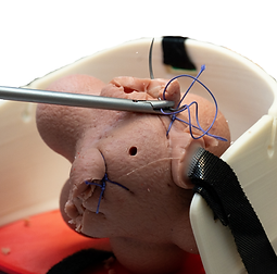
Model Validation at ORSI
Mirai Surgical collaborates with leading surgical training centers to validate its simulation models under real procedural conditions.
At ORSI, expert surgeons performed a step-by-step robotic myomectomy procedure using the Mirai Surgical model to assess anatomical accuracy, tissue-like texture, and overall realism.
This validation confirms the model’s suitability for standardized training, enabling reproducible gynecology education in a highly realistic and controlled environment.

